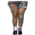
Mary has recently undergone a full knee replacement. Watch the video of how her arthritis and knee deformity have been corrected with surgery.
Dialog from Mary's Knee Replacement Video:
Dialog from Video:
Mary has recently undergone knee replacement surgeryknee replacement surgery. This x-ray was taken two weeks following her procedure.
Let’s see what Mary’s knee looked like before surgery. Her left knee has a significant knock-knee deformity. Her left hip is even turned inward to try to compensate for her arthritic knee. Here is the x-ray of Mary’s whole left leg. As I draw a straight line from her hip to her ankle, notice how Mary’s knee sits significantly to the inside of this line. Before she had developed arthritis, Mary’s knee was positioned directly through this line. The years of arthritisarthritis have caused her knee to move out of normal alignment.
Here is a closer look at Mary’s knee before surgery. When, I apply her x-ray on top of her leg, her arthritis starts to become more apparent. She has significant joint space narrowing on the outside of her knee, leading to her knock-knee deformity.
Let’s review Mary’s standing x-ray in more detail. The top bone is the thigh or femurfemur bone, the bottom bone is the shin or tibiatibia bone. The knee is a hinge joint where these two bones meet together. In Mary’s knee there is a loss of cartilage with development of bone spurs called osteophytes. See what happens when Mary bends her knee slightly and we take another x-ray. Watch by the arrow on the outside of her knee. The arthritis becomes even more pronounced. The space between the bones has been completely obliterated with severe bone on bone contact and worsening of the angular deformity of the knee. Let’s watch this again.
Mary tried multiple conservative measures before considering knee replacement. These included the use of a bracebrace, cortisone injectionscosrtisone injections, hyaluronic acid injectionshyaluronic acid injections and physical therapyphysical therapy. Here is Mary’s leg two weeks after surgery. Her leg is still swollen, and she has some normal bruisingbruising. Let’s move forward to three months after surgery. Her incisionincision is well healed and her swelling has resolved. Mary can now walk without pain, and the surgery has straightened her knee. Let’s review her pre-operative picture once again. The difference between the before and after is dramatic.
Now let’s look at Mary’s full length post-operative x-ray laid on top of her leg. Her knee now sits in the straight line that is drawn from her hip to her ankle. Here is a closer look at her knee prosthesisknee prosthesis. I can then place the post-operative x-ray on top of her leg. Let’s review the x-ray in more detail. This is the new metal component on the femur. Here’s the new metal component on the tibia. Notice how the space on the inside and outside of the knee is now equal. That space is occupied by a special plastic called polyethylene.
Here is the side view Mary’s leg when she is lying down two weeks after surgery. Let’s place the x-ray of the new knee replacement on her leg. Notice that the swelling prevents her knee from getting completely straight. Now we can look at the standing side view of her leg after her healing has completed. Here is Mary’s x-ray layed on top. Her motion after knee replacement surgerymotion after knee replacement surgery has significantly improved and she can now stand with her knee fully extended.
Mary’s knee no longer limits her life. Her knee function has significantly improved and she is able to return to her favorite activities including bike riding and golf after knee replacementbike riding and golf after knee replacement. Mary’s surgeon has been with her every step of the way, just as My Knee Guide will be with you.


